

What Is Needed To Register A Car In Ct
- What is a computed tomography (CT) scan?
- How does CT piece of work?
- When would I get a CT scan?
- What is a CT contrast agent?
- Are in that location risks?
- What are examples of NIBIB-funded projects using computed tomography?
What is a computed tomography (CT) browse?
The term "computed tomography", or CT, refers to a computerized 10-ray imaging procedure in which a narrow beam of x-rays is aimed at a patient and chop-chop rotated around the body, producing signals that are candy by the machine'south computer to generate cross-sectional images—or "slices"—of the body. These slices are chosen tomographic images and contain more detailed data than conventional x-rays. Once a number of successive slices are collected past the machine's figurer, they can exist digitally "stacked" together to grade a three-dimensional image of the patient that allows for easier identification and location of basic structures as well equally possible tumors or abnormalities.
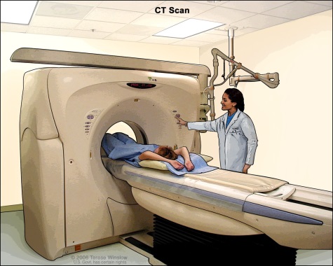
How does CT work?
Unlike a conventional x-ray—which uses a fixed 10-ray tube—a CT scanner uses a motorized x-ray source that rotates around the circular opening of a donut-shaped structure chosen a gantry. During a CT scan, the patient lies on a bed that slowly moves through the gantry while the x-ray tube rotates around the patient, shooting narrow beams of x-rays through the body. Instead of film, CT scanners use special digital ten-ray detectors, which are located directly opposite the 10-ray source. Equally the x-rays exit the patient, they are picked up past the detectors and transmitted to a computer.
Each fourth dimension the x-ray source completes one full rotation, the CT computer uses sophisticated mathematical techniques to construct a 2D image slice of the patient. The thickness of the tissue represented in each image slice tin vary depending on the CT car used, but usually ranges from 1-10 millimeters. When a full slice is completed, the image is stored and the motorized bed is moved forward incrementally into the gantry. The 10-ray scanning process is then repeated to produce another epitome slice. This process continues until the desired number of slices is nerveless.
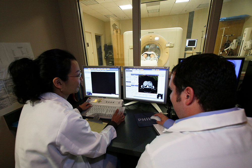
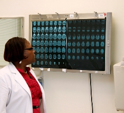
Image slices tin either be displayed individually or stacked together by the computer to generate a 3D image of the patient that shows the skeleton, organs, and tissues also as any abnormalities the doc is trying to place. This method has many advantages including the ability to rotate the 3D image in infinite or to view slices in succession, making information technology easier to find the exact place where a problem may be located.
When would I become a CT scan?
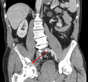
Source: James Heilman, Chiliad.D., [CC-BY-SA-three.0]
CT scans can be used to place disease or injury within various regions of the body. For case, CT has become a useful screening tool for detecting possible tumors or lesions within the abdomen. A CT scan of the heart may be ordered when various types of heart disease or abnormalities are suspected. CT can also exist used to epitome the head in order to locate injuries, tumors, clots leading to stroke, hemorrhage, and other conditions. It can prototype the lungs in order to reveal the presence of tumors, pulmonary embolisms (blood clots), excess fluid, and other conditions such as emphysema or pneumonia. A CT scan is particularly useful when imaging complex bone fractures, severely eroded joints, or bone tumors since it ordinarily produces more than particular than would exist possible with a conventional 10-ray.
What is a CT contrast agent?
As with all x-rays, dense structures within the body—such as bone—are hands imaged, whereas soft tissues vary in their power to stop x-rays and, thus, may be faint or hard to run into. For this reason, intravenous (IV) contrast agents have been developed that are highly visible in an x-ray or CT browse and are safety to use in patients. Contrast agents incorporate substances that are meliorate at stopping x-rays and, thus, are more visible on an ten-ray image. For case, to examine the circulatory organisation, a contrast agent based on iodine is injected into the bloodstream to assistance illuminate blood vessels. This type of test is used to look for possible obstructions in claret vessels, including those in the middle. Oral dissimilarity agents, such every bit barium-based compounds, are used for imaging the digestive system, including the esophagus, stomach, and GI tract.
Are there risks?
CT scans tin diagnose perchance life-threatening conditions such every bit hemorrhage, blood clots, or cancer. An early diagnosis of these conditions could potentially be life-saving. However, CT scans use x-rays, and all x-rays produce ionizing radiations. Ionizing radiation has the potential to cause biological furnishings in living tissue. This is a risk that increases with the number of exposures added upwards over the life of an individual. Nonetheless, the hazard of developing cancer from radiation exposure is generally small.
A CT scan in a pregnant woman poses no known risks to the infant if the surface area of the body being imaged isn't the abdomen or pelvis. In general, if imaging of the abdomen and pelvis is needed, doctors prefer to employ exams that do non use radiation, such as MRI or ultrasound. Even so, if neither of those tin can provide the answers needed, or in that location is an emergency or other fourth dimension constraint, CT may exist an acceptable culling imaging option.
In some patients, contrast agents may cause allergic reactions, or in rare cases, temporary kidney failure. Iv contrast agents should not be administered to patients with abnormal kidney role since they may induce a farther reduction of kidney function, which may sometimes become permanent.
Children are more sensitive to ionizing radiation and have a longer life expectancy and, thus, a higher relative risk for developing cancer than adults. Parents may want to inquire the technologist or doctor if their machine settings have been adjusted for children.
What are examples of NIBIB-funded projects using computed tomography?
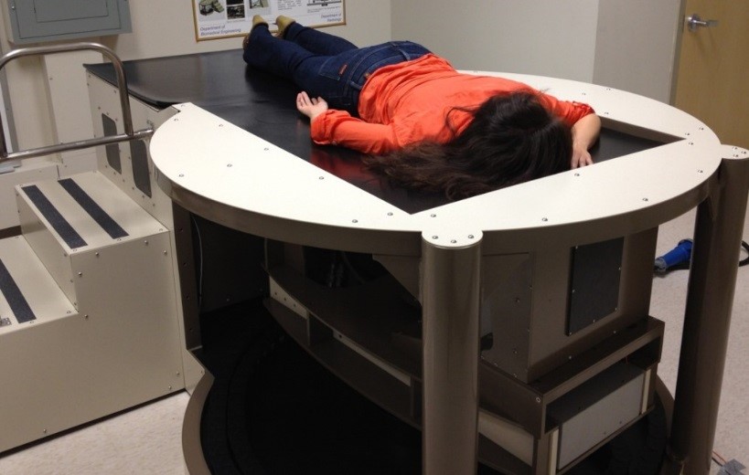
Credit: John Boone, UC Davis
Dedicated Breast CT Scanner: NIBIB is funding enquiry for development of a dedicated breast CT scanner that allows the breast to be imaged in 3D and could help radiologists discover hard-to-find tumors. The scanner produces a radiation dose comparable to that of a standard x-ray mammogram and doesn't require compression of the chest. In this breast CT scanner, a woman lies prone in a peculiarly designed large table with her breast suspended in a special opening in the scanning bed. The scanner rotates around the chest, without passing through the chest, thus reducing the radiation that would exist delivered to the chest in a conventional CT scanner. Mind to a podcast virtually the scanner.
Reduction in Radiation from Routine CT Scans:NIBIB put out a call for researchers to submit groundbreaking ideas that will help to radically decrease the amount of radiation used in CT scans. Five new projects are underway from this new funding opportunity, representing artistic, innovative, interdisciplinary approaches that would not take been funded otherwise. You can read more well-nigh them below:
Customized imaging
Web Stayman, Johns Hopkins University
The amount of radiations required for a CT scan depends on a number of variables, including the size of the patient, the part of the body being scanned, and the diagnostic task at hand. For example, smaller patients require less radiation than larger patients, and scanning a denser function of the trunk, such every bit soft tissue near the pelvis, requires more radiation than scanning the lungs. In addition, diagnostic tasks that crave high image clarity, such as locating a faint tumor, mostly require more radiation. The goal of this project is to modify both the hardware and software of mod CT systems so that the device can adapt the shape, position, and intensity of the 10-ray beam to the specific imaging scenario. The inquiry leverages patient-specific anatomical models and mathematical models of imaging performance to straight 10-rays where they are needed and, consequently, to avoid or to limit ten-ray exposure where it is not needed. This will assist maximize imaging performance for specific diagnostic tasks while minimizing radiations exposures.
Constructing tools for researchers
Cynthia McCollough, Mayo Dispensary
The goal of this work is to develop resources that enable the research community to easily create and compare new approaches to reducing radiation dose of routine CT scans without compromising diagnostic accuracy. So far, this has entailed creating a library of raw information from patient CT scans that researchers can manipulate to examination new approaches, and developing calculator-based methods for evaluating new approaches, and then that researchers don't take to rely on radiologists, which tin can be costly and time consuming. Using these assets, researchers have demonstrated that there is considerable potential for radiation dose reduction in CT exams of the abdomen, which are among the highest dose CT exams in mutual clinical use.
Faster processing
Jeffrey Fessler, University of Michigan
To reduce radiation yet withal produce good quality CT images, more sophisticated methods are needed to process the raw data from the CT system. Those advanced methods, called paradigm reconstruction algorithms, can require undesirably long computing times, so they can be used only for some patients currently. The goal of this project is to develop algorithms that are fast enough to let depression-dose CT imaging to be used for every patient.>
An integrated approach
Norbert Pelc, Stanford Medical School
At every stage in the design of CT scanners, there are opportunities to make changes that reduce radiation dose. Because these changes are inter-related, the goal of this project is to take an integrated approach, exploring approaches such as modifying the photon counting detector (the part of the CT scanner that detects x-rays), dynamic x-ray illumination (adjusting the amount of radiation used throughout the elapsing of a browse), and prototype reconstruction methods. These will be tested using a table acme experimental organisation. The researchers believe that these combined strategies can lead to as much as 80% reduction in radiations dose compared to today's typical systems, and too enable higher resolution images.
SparseCT
Ricardo Otazo and Daniel Sodickson, New York University School of Medicine
Investigators at New York Academy School of Medicine, Brigham and Women'southward Infirmary, and Siemens Healthineers are working together to develop a new ultra-depression-dose CT technique called SparseCT. The cardinal idea backside SparseCT is to block virtually of the Ten-rays in a CT browse earlier they attain the patient, but to practice and then in a way that preserves all the essential image information. The approach combines a new x-ray blocking device with the mathematics of compressed sensing, which allows images to exist reconstructed from reduced datasets. Pinch sensing can be likened to filming a film with a very fast, simply low-pixel camera and so using math to convert the image to high-definition quality.

Source: https://www.nibib.nih.gov/science-education/science-topics/computed-tomography-ct
Posted by: allenleareved.blogspot.com
0 Response to "What Is Needed To Register A Car In Ct"
Post a Comment